
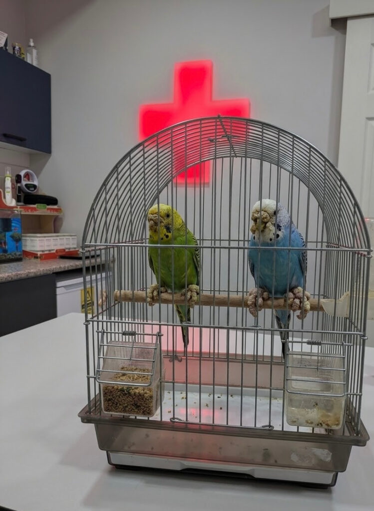
When dryness is observed at the corners of a budgerigar's beak or on its feet, the question "is it mites or fungus?" frequently arises. Mite and fungal infections are caused by different agents and are not treated with the same medication. White, porous scabs may suggest mite infestation, but it is not possible to make a definitive diagnosis based solely on a photograph. If the beak is changing shape, the bird is having difficulty breaking food, or if it cannot use its foot comfortably, examination should not be delayed.
While the term 'mites' may evoke severe itching, this condition can present with mild itching or may even go unnoticed. Even if the bird is not scratching, the cause of the scabs on the beak and feet should be investigated.
What are scaly face and scaly leg mites?
In budgerigars, this condition is usually caused by a microscopic mite called Knemidokoptes pilae. The term Knemidocoptes may also be encountered in literature. The mite lives in the superficial layers of the skin that contain keratin; the resulting tissue alteration can gradually create a scabby and porous appearance.
In common parlance, this appearance is sometimes called "beak fungus." However, mites are not a fungal disease. Misnaming the cause can lead to the use of ineffective products and delays in the proper treatment. Associated feather changes are also examined; there are various causes of feather disorders in birds.
What do mite infestations look like in budgerigars?
Initially, a light whiteness may be observed at the corner of the beak or in the area known as the cere, where the nostrils are located. Over time, the scabs may become more pronounced and spread to other areas beyond the beak.
Changes that should be monitored include:
- Whitish scabs on the beak edge, cere, or around the eyes,
- Spongy or flaky surfaces containing small holes,
- Inequality and distortion in beak growth,
- Thickening of the skin on the feet and toes, with scales becoming more pronounced,
- In advanced cases, difficulty in breaking food or using the foot.
Not all symptoms need to be present at the same time. Even changes that start in just a small area warrant an examination. A bird going to its feeder does not alone indicate that it is eating adequately; whether it is actually breaking and consuming the food is also important.
Can there be a fungal infection on the beak and feet?
Fungal infections affecting the skin can occur in birds. Yeasts like Candida and some other fungi can cause skin diseases; a fungal infection may also develop in the keratin layers of the beak. However, it is not correct to assume that every scaling on the budgerigar's foot or every whiteness around the beak is a "fungus." Skin irritation, crusting, and wound appearance can resemble each other in different diseases.
The outer surface of the beak and the inside of the mouth are evaluated separately. If there are white plaques inside the mouth, difficulty swallowing, or vomiting, diseases of the mouth and digestive system should be investigated. Candida is more associated with areas such as the mouth, esophagus, and crop; looking at a scab on the outer beak cannot lead to a diagnosis of crop fungal infection. This condition is also discussed in our article on crop fungal infection in birds.
Compromised skin integrity, issues in the bird's overall health, and the use of certain medications can predispose to infection. During the examination, previous antibiotics and creams applied will therefore be questioned. Suspicions of fungal infections are not sufficient grounds for selecting treatment without proper samples.
Is every beak scab or elongation due to mites?
No. In particular, in adult female budgerigars, the cere can appear brown and thick due to the effects of reproductive hormones. This change is not the same as the porous crusting seen in mites. Nonetheless, a formation that narrows the nostrils, changes rapidly, or differs from the usual appearance should be checked.
Deformities in the beak may also be related to other diseases or prior injuries. Scaling on the foot and a red, painful wound on the sole of the foot should also be evaluated separately. The distinction in sole lesions is important, as discussed in our article on foot sole lesions and pododermatitis in birds. Choosing a product without seeing the location and structure of the crusting may overlook a problem other than mites.
Can it spread to other birds?
Close contact is important for the transmission of mites; birds sharing the same environment should be evaluated together. However, not every bird that comes into contact will necessarily develop symptoms at the same time. A second bird at home having a normal-looking beak does not mean it does not need evaluation.
Inform the veterinarian if a new bird has been added, the duration the birds have been living together, and the products used previously. An examination for contact birds, distinguishing features, and if necessary, a treatment plan should be determined by the veterinarian. Precautions for contact birds and the environment regarding fungal infections vary depending on the identified agent. Do not apply the same dosage of medication given to one bird to another.
How do veterinarians distinguish mites from fungal infections?
The veterinarian examines the beak, cere, inside the mouth, and feet; they assess the location of the lesions, the function of the beak, and the overall health of the bird. In cases of suspicion of mites, demonstrating the mites in samples taken from the appropriate areas supports the diagnosis.
In suspected fungal cases, a cytological examination of skin or lesion samples can be performed, meaning that cells and microorganisms are evaluated under a microscope. If necessary, a fungal culture or tissue examination may be planned for persistent or deep lesions. The presence of a microorganism in the sample is interpreted together with the appearance of the lesion and other findings. Samples are not taken from every bird using the same method.
Why are mite and fungus treatments different?
For mites, appropriate antiparasitic treatment is applied to the patient. If a fungal infection is confirmed, antifungal treatment is selected based on the agent and the condition of the affected tissue. A product used on the skin cannot replace a medication for the mouth or crop. Accompanying care and dietary issues also need to be addressed. Even if the scabs decrease, do not discontinue the treatment on your own; do not skip the repeat applications specified in the prescription. During the follow-up examination, the status of active lesions and the function of the beak are reassessed. In the use of patient-specific medications in birds, the concentration of the product and the route of administration are as important as the given amount.
In advanced beak deformities, in addition to parasitic treatment, the function of the beak should also be evaluated. The elimination of parasites does not mean that all previously formed shape changes will immediately correct. Some birds may require long-term monitoring of the beak.
What should you pay attention to at home until the examination?
Do not pull off crusts, do not cut the beak, and do not try to peel off the thickened skin on the foot. Do not use anti-mite creams made for humans, random parasite sprays, or drops meant for other animals. Applying oil can change the appearance of the crusts but will not provide the correct treatment for mites or fungus.
Ensure cleanliness of the cage, feed, and water bowl. Consult your veterinarian on which materials can be cleaned and which need to be replaced; do not spray cleaning products on the bird. Bringing the packaging of existing products and photos showing the progress of the change to the examination can be helpful.
If the bird cannot eat, shows significant lethargy, bleeds, cannot stand on its foot, or has difficulty breathing, do not delay. If there is a ring on the leg, swelling and constriction around it should also be evaluated without delay; do not attempt to cut the ring at home. If the bird shows open-mouth breathing or tail movement with breathing, pay additional attention to signs of respiratory distress.
When seeking help for beak and foot issues, you can prepare a list of products used and photos of the cage. You can find information about the evaluation process on our Bird Veterinarian Examination page.
Frequently Asked Questions
Are mites and fungus the same disease?
No. Mites are a parasitic disease caused by microscopic mites; the agent in fungal infections is different. They can be confused due to their ability to cause changes in similar areas. Antiparasitic medication cannot be used interchangeably with antifungal medication.
My budgerigar is not scratching; can it still have mites?
Yes. In beak and foot mites, itching is not always prominent. The location, structure, and changes over time of the scabs may be more indicative than the behavior of scratching.
Can I use human cream for beak fungus or foot fungus?
No. The product's content affects how the bird's beak receives the medication and the safety of the applied area. It should first be determined whether there are mites, fungus, or another skin issue; the product and method of use should be selected by the veterinarian.
Does the beak immediately recover after mite treatment?
Not always. Even if the active parasitic problem is controlled, previously developed deformities may persist. The bird's ability to break food and the manner in which the beak elongates are monitored in follow-ups.
Does simply cleaning the cage cure the disease?
No. Maintaining cleanliness and care is important, but it does not replace the necessary treatment for mites or confirmed fungal infections. The plan for contact birds should also be evaluated separately.
How long does mite and fungus treatment take?
The duration depends on the diagnosis, the extent of lesions, damage to the beak and feet, and the response to treatment. Improvement in the appearance of scabs does not mean that the treatment is complete; your veterinarian will determine the follow-up date.
This content is for general informational purposes; it does not replace a veterinarian examination.

